Digitizing Hope: Preserving a Species on the Brink of Extinction

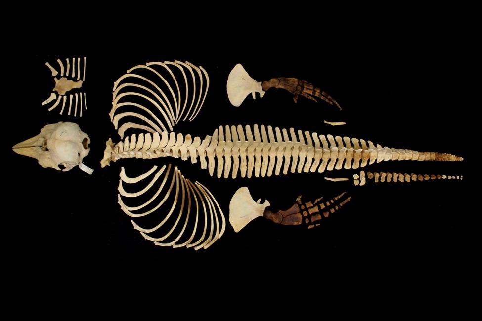
Full skeleton of a very rare vaquita specimen from the 1960s. The completed scans, which required approximately 165 hours, resulted in a total of three terabytes of data. (Photo credit: Jamie Knaub, Florida Atlantic University)
The vaquita, which means “little cow” in Spanish, is the world’s smallest porpoise and most endangered marine mammal. They also have the smallest range of any marine mammal; about 1,500 square miles within the northern Gulf of California. Since 1997, vaquitas have experienced a dramatic population loss from about 600 individuals to an estimate of less than 10 animals to date. At this current rate, vaquitas are expected to become extinct imminently.
The vaquita’s decline is caused by entanglement in illegal gillnets used to fish totoaba, an endangered species prized for its swim bladder. Despite a gillnet ban and conservation efforts, the illegal totoaba trade continues due to organized crime and poaching. Conservation actions include global awareness campaigns, removing gillnets, monitoring vaquitas and combating poaching, but attempts by other organizations to protect vaquitas in captivity have been unsuccessful.
While hope for the recovery of vaquitas is vital, immediate action to preserve this endangered species is even more crucial. One powerful step toward safeguarding their future lies in the digitization of the vaquita anatomy.
Florida Atlantic University , in collaboration with the San Diego Natural History Museum and SeaWorld San Diego , is playing an important role in this preservation. Using state-of-the-art, high resolution micro-CT scanning in the FAU High School Owls Imaging Lab, researchers have scanned a full skeleton of a very rare vaquita specimen.
“We are delighted to collaborate with likeminded organizations to make our collections as useful and accessible as possible,” said Phil Unitt, curator of birds and mammals at the San Diego Natural History Museum. “A complete skeleton of a vaquita is an extremely rare specimen, so we’re thrilled to learn its replica will be available to the public.”
The skeleton, on loan from the San Diego Natural History Museum to SeaWorld San Diego, is thought to be one of, if not the only, full vaquita skeleton available in the United States. The skeleton was donated to the museum in 1966. The objective of scanning this rare specimen for display purposes is to facilitate the creation of replicas to be commercially available to further education and conservation efforts of this critically endangered species.
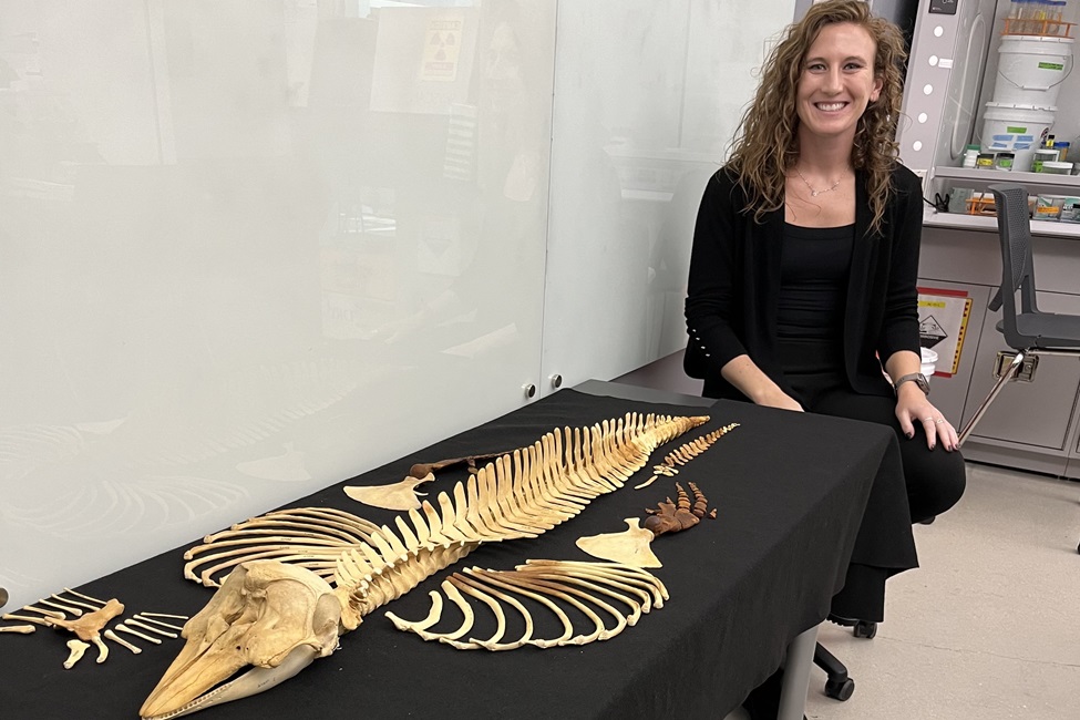
“The specimen we scanned was an adult female vaquita and a rarity so significant that it could not be shipped and required careful escort for transportation,” said Jamie Knaub, imaging lab assistant in the FAU Lab Schools’ Owls Imaging Lab and a Ph.D. candidate in the FAU Department of Biology within the Charles E. Schmidt College of Science. “In August, I traveled to San Diego to acquire the skeleton from SeaWorld and personally transported it back to Florida as carry-on luggage. The specimen was housed in the Owls Imaging Lab for three months during the scanning process. The completed scans, which required approximately 165 hours, resulted in a total of three terabytes of data. I returned the skeleton to SeaWorld for safekeeping in early December.”
Knaub has been working with Brittany Aja Dolan, pathology and research associate at SeaWorld San Diego, who spearheaded the project. Knaub previously collaborated with Dolan who provided her with thresher shark vertebrae from a stranded shark in California for her graduate research, which was used in her first dissertation chapter and resulted in publication in the journal of the Royal Society. Knaub published the paper with Marianne E. Porter, Ph.D., senior author and an associate professor, FAU Department of Biological Sciences; and Tricia Meredith, Ph.D., director of research for FAU’s on-site lab schools, A.D. Henderson University School and FAU High School, and an assistant research professor in FAU’s College of Education.
“The imminent extinction of the vaquita is a sobering reminder of the impact that humans can have on the wildlife and environment, said Dolan. “According to genetic studies there is hope for their successful recovery, and through this unique multifaceted collaboration, we have immortalized a one-of-a-kind skeleton. We hope that by creating replicas, which will be available worldwide, and hopefully on display at SeaWorld San Diego in the near future, everyone will have the opportunity to learn about the world’s most endangered marine mammal and what we can all do to help.”
Initial CT scans of this rare vaquita specimen were completed by the San Diego Zoo but were not sufficient resolution for replication. Dolan contacted Knaub about employing FAU’s micro-CT scanner to obtain high resolution scans of the skeleton. The San Diego Natural History Museum and SeaWorld San Diego have given Knaub permission to use the vertebral scans of the vaquita in her dissertation research.
“At the rate that vaquitas are disappearing, it’s extremely important to preserve as much about this species as we can,” said Knaub. “They are very elusive and not many physical specimens from this species exist.”
The 3D scans of this vaquita skeleton will be hosted on MorphoSource, a publicly accessible data repository dedicated to housing image data that represents physical objects of our world. The scans will be available for download and used for education, outreach, and research purposes. Additionally, SeaWorld San Diego will be working with Bone Clones to produce full replicas of the vaquita skeleton for education.
“Imaging such a rare specimen is important because the digital representation of this individual such as photos, scans, and 3D mesh files will persist long after the last living vaquita is gone,” said Knaub. “Importantly, digitizing the skeleton and making the data openly available to other researchers and the public significantly enhances accessibility, providing broader opportunities for collaboration and research.”
The FAU Owls Imaging Lab is a one-of-a-kind research laboratory that provides students access to cutting-edge equipment to work on high-level research projects, including cancer treatment research, vaccine development, and prosthetic creation, among others. Students can research some of the world’s most challenging problems at an early age and can share that research and publish it in peer-reviewed journals. The lab includes a micro computed tomography scanner; scanning electron microscope; histology suite; inverted compound microscope; and stereoscope and is available to students and faculty at A.D. Henderson University School, FAU High School, and all FAU colleges.
“The primary aim of our open-access research hub is to create a dynamic environment that promotes meaningful collaborations between our students and university mentors. By providing opportunities for hands-on teaching, innovative demonstrations, experimentation, and robust data collection, the hub seeks to enhance the educational experience and advance research excellence,” said Meredith. “These collaborations not only deepen students’ understanding of scientific methodologies but also support the creation of impactful, high-quality publications and presentations that contribute to their academic and professional growth. Through this initiative, we strive to build a community of scholars dedicated to advancing knowledge and addressing real-world challenges.”

Jamie Knaub pictured with the full skeleton of a very rare vaquita specimen from the 1960s. (Photo credit: Tricia Meredith, Ph.D., Florida Atlantic University)

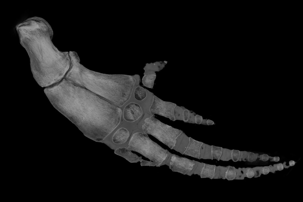
Volume rendering of the flipper of a very rare vaquita specimen from the 1960s. (Photo credit: Jamie Knaub, Florida Atlantic University)

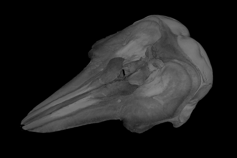
Volume rendering of the skull of a very rare vaquita specimen from the 1960s. (Photo credit: Jamie Knaub, Florida Atlantic University)
-FAU-


